
Tag season is about to start. North City Physiotherapy are the go to physio’s to help you get through your season. Whether its a hammy strain, pulled calf, or you have rolled ankle we can help you. Careful pulling those flags off, fingers! We can offer 15% of all physiotherapy services for PC tag players. Just let us know you play in the PC Tag competition and we will sort you out. Don’t delay book now!
Back pain? Physiotherapy can help.
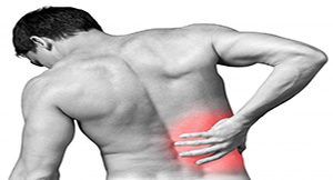
Check how how physio can help with back pain:
Body weight circuits Part One – The humble press up

Welcome to the first of a series of exercise based newsletters from me that will make up a full body weight circuit that can be done without equipment, vast amounts of space or, lengthy periods of time.
I was first introduced to body weight circuits as a scrawny middle distance runner trying to build a bit of strength. As hard as I found them I can only say that they were effective and, they are exercises that not only have passed the test of time but form the mainstay of the approach that I now take to physiotherapy and rehabilitation. My ethos on exercise is that I want to see the benefits not only within the exercise setting but outside in any individuals day to day life. Consequently exercises I use mimic functional movement patterns that are used every day by us all, for example pushing and pulling movements or squatting and lunging movements or twisting and rotational movements. Therefore today we will be looking at the humble press up as an exercise that will mimic pushing actions.
First up what does a press up work, well done correctly it will challenge your whole body but specifically core, pectoral and tricep muscles. Let me explain further. Your core muscles are your foundation, they give you the base to work from without a sturdy core any functional movement will be less effective. What we know about these muscles is that they will fire and activate before any other skeletal muscle regardless of what the movement is – I could be pushing a door open or standing up from a chair and my core will fire first. The pectorals are a made up of two muscles – Pectoral major and Pectoral minor that principally influence movements of the shoulder. The tricep is a large muscle on the back of your arm. It primarily extends or straightens the elbow but, because it partly attaches on to the scapular (shoulder blade) it does have a role in shoulder movements.
Now knowing what a press up does the next question is how do we do perform one. The classic version of a press up starts with your hands beneath your shoulders, face down and with the next point of contact being your toes. The movement is to press up and away from the floor and then lower back down, elbows remain tucked in to your sides and your back remains straight without any increased curve in your lower back.
e99c39f7 013f 41e0 a322 66071854e6ab
What is important to know however is that this is not the only way to perform a press up. As with any exercise if you shorten it, destabilise it, start it from a different position you can influence it so, here are my rough progressions. Remember though what is key is that the variety of press up that is selected should challenge you over the sets and repetitions you select, for example if I wanted to do three sets of ten repetitions I want to be struggling to complete the last couple of repetitions, if I come nowhere near that or could do an extra three sets I have not selected the right variation, so here are my progressions:
851d5897 0476 4288 be9c 1217732404a8
psuh up 3
Good luck and watch out for the second instalment of the body weight circuit.
CHRISTMAS AND NEW YEAR PERIOD APPOINTMENTS

Hey Everyone, just to let you know we are open during the Christmas and New Year period on the non public holiday period. Please don’t hesitate to contact us should you need an appointment, we have 3 physiotherapists working throughout this time.
Exercise technology

At North City Physiotherapy we pride ourselves in offering our patients the latest technologies to help you achieve your outcomes. To create your tailored exercise programs we use a brand new app called PhysiApp. PhysiApp lets you complete your prescribed exercise program by following crystal-clear, narrated exercise videos. PhysiApp is completely free to download from the App store and Google Play store or can be accessed via your browser. Built-in reminders help you to stay on track towards a better you! See some of the below examples of the great content:
Medical Imaging

By James Flannery – Physiotherapist
In this newsletter, we’ll take a look at the three primary types of medical imaging that you might come across while under the care of your physiotherapist. I’ll look to outline what each is, what it shows us, and why it’s used.
First up, X-rays:
Most people are familiar with what an x-ray looks like, but I regularly have patients who aren’t really sure what they’re used for.
An x-ray machine works by firing x-rays, a type of electromagnetic radiation, through the target area, toward a detector plate on the other side. While this sounds somewhat scary at first, rest assured that the x-rays pass harmlessly through most tissues in the body, and are deflected away by harder tissue such as bone. The x-rays that hit the detector plate darken it, and image that is left behind after an x-ray is the shadow of the structures that could not be penetrated by the x-rays.
X-rays are an effective tool when there’s a need to check for bony injury; after a bad ankle injury, or a fall onto an outstretched wrist for instance. We can also use x-rays to check for some types of lung conditions, as fluid or other denser materials in the lungs shows up as lighter areas than air-filled areas of the lung.
X-rays do not show softer tissues like muscles and ligaments, and so can’t be used to directly diagnose damage to these structures; however, if an x-ray doesn’t show any bony injury, we can rule that out as a potential source of pain, which can help narrow down the diagnosis.
The second most commonly used imaging technique is diagnostic ultrasound. The most familiar use of an ultrasound scan to most people will be during pregnancy, during which a doctor can use ultrasound to look at a growing foetus and ensure that it’s healthy.
The same technique can be applied on other areas in order to check for things like muscle and ligament tears. The ultrasound probe emits high-frequency sound waves, which echo back off structures within the body, with different tissues reflecting at different levels. By picking up these echoes, the machine can create an image of the area within the body.
Ultrasound is a useful tool for early diagnosis of soft tissue injuries; it can be useful to tell whether something like a shoulder injury can be managed purely through physiotherapy, or whether the level of injury is such that it might need referral to a specialist.
One of the main limitations of ultrasound is that it is difficult to pick up injuries that are particularly deep, or that are surrounded by bony structures. Things like anterior cruciate ligament injuries in the knee can’t be reliably viewed by ultrasound.
The last main type of imaging is MRI, or magnetic resonance imaging. MRI is significantmy more complex than the other two forms of imaging, and involves creating a powerful magnetic field into which the patient is placed, then detecting the changes in vibrations of atoms throughout the patient’s body in response to the magnetic field; this is done by using concept known as nuclear magnetic resonance. Note that ‘nuclear’ in this sense refers to the core of the atoms; the process is actually free of any potentially harmful radiation.
The machines used for MRIs are very complex, very large, and very expensive. Because of this, and a few other reasons, there are limitations on who is able to refer patients for MRI scans. Neither physiotherapists nor GPs are able to refer directly for these scans, and will instead refer patients who might need them to a specialist, often an orthopaedic surgeon or a sports doctor.
An MRI scan gives us a very clear picture of all structures in the area scanned; ligaments, muscles, bones, nerves etc, and, for most things, is the best and most accurate form of imaging that we have available. MRIs are generally reserved for more serious injuries and/or for injuries that might need to be managed surgically.
Hopefully this has given a little more insight into an otherwise confusing area of medicine. If you have any questions about any imaging that’s been done for your injuries, ask your physiotherapist, and they’ll likely be happy to clear things up for you.